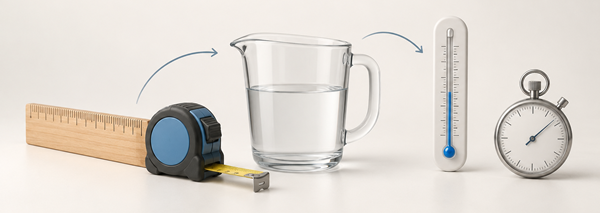
Convert microseconds to seconds when examining ultrasound echoes, electronic signals and other extremely short events. This microseconds to seconds converter divides the microsecond value by 1,000,000 and displays the result instantly. Explore the formula, common values and the educational story of how echoes returning in microseconds help an ultrasound scanner build images that can be viewed over several seconds.
Microsecond to Second Conversion Formula
The conversion formula is:
1 µs = 0.000001 s
Or simply:
µs × 0.000001 = s
This conversion uses fixed seconds-based factors: 1 µs = 0.000001 s and 1 s = 1 s.
Microsecond to Second Conversion Table
| Microseconds (µs) | Seconds (s) |
|---|---|
| 1 µs | 0.000001 s |
| 5 µs | 0.000005 s |
| 10 µs | 0.00001 s |
| 50 µs | 0.00005 s |
| 100 µs | 0.0001 s |
| 500 µs | 0.0005 s |
| 1,000 µs | 0.001 s |
| 5,000 µs | 0.005 s |
| 10,000 µs | 0.01 s |
| 100,000 µs | 0.1 s |
| 500,000 µs | 0.5 s |
| 1,000,000 µs | 1 s |
To convert microseconds to seconds, divide the number of microseconds by 1,000,000. One second contains exactly 1,000,000 microseconds.
How Microsecond Echoes Build an Ultrasound Image in Seconds
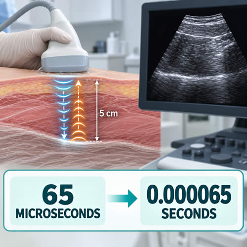
During a medical ultrasound examination, a clinician moves a transducer across the patient's skin while images appear on a nearby screen. The movement of an organ or blood flowing through a vessel may be displayed in real time, but every visible image begins with sound waves travelling through the body and returning as echoes.
An individual echo can complete its journey in microseconds. The scanner sends and receives many pulses, processes their timing and strength, and combines the information into images that can be viewed over a period of seconds.
The Transducer Sends a Sound Pulse
An ultrasound transducer contains materials that can convert electrical energy into high-frequency sound waves. The same transducer can detect echoes and convert them back into electrical signals for processing.
A layer of gel is normally placed between the transducer and the skin. The gel helps prevent air pockets from interfering with the transmission of sound into the body.
As a pulse travels through the body, some of its energy is reflected at boundaries between different tissues. These returning reflections are the echoes used by the scanner. The system considers both the echo's return time and its strength when producing an image.
The timing principle behind this traces back to a much older technology. Medical pulse-echo imaging grew directly out of SONAR, developed for underwater detection, and the technique's modern medical use dates to work by Scottish obstetrician Ian Donald, whose 1958 publication on diagnostic ultrasound in obstetrics helped establish it as a clinical tool. The same timing principle — measuring how long a pulse takes to return — still underlies every ultrasound scan performed today.
The National Institute of Biomedical Imaging and Bioengineering explains that the scanner uses the speed of sound and the time taken for each echo to return when calculating the distance to a tissue boundary.
A Microsecond Journey into the Body
Medical pulse-echo systems commonly perform their calculations using an assumed sound speed of approximately 1,540 metres per second in soft tissue. Because the pulse must travel to a structure and return to the transducer, the calculation uses the complete round-trip distance.
Imagine a simplified example in which a tissue boundary is 5 centimetres beneath the transducer. The sound travels 5 centimetres into the body and another 5 centimetres back:
Round-trip distance = 0.05 m × 2 = 0.10 m
Dividing the distance by the assumed sound speed gives an approximate echo time:
0.10 m ÷ 1,540 m/s ≈ 0.0000649 seconds
To express this result in microseconds:
0.0000649 s × 1,000,000 ≈ 64.9 µs
The echo therefore completes its simplified round trip in approximately 65 microseconds. Actual ultrasound behaviour is more complex because sound speed and reflection characteristics vary among tissues.
Converting the Echo Time into Seconds
A microseconds to seconds converter helps place the echo delay on a familiar time scale. To convert microseconds to seconds, divide the microsecond value by 1,000,000:
Seconds = microseconds ÷ 1,000,000
For the approximate echo time in this example:
65 µs ÷ 1,000,000 = 0.000065 seconds
The two values describe the same interval. Microseconds are more practical for examining individual echoes, while seconds are more useful for describing a complete sequence or the visible passage of time.
Many Echoes Create One Image
One returning echo does not create a complete anatomical image. The scanner repeatedly transmits pulses along multiple paths and measures echoes returning from different depths. It processes those signals and assigns image positions and brightness levels based on the detected information.
This process is repeated rapidly to produce successive image frames. Individual measurements may be separated by microsecond-scale intervals, while the resulting moving image can be observed for several seconds or longer during an examination.
The US Food and Drug Administration notes that ultrasound images are produced from reflected sound and the time taken for waves to travel through the body. It also explains that ultrasound can display movement in real time and does not use the ionising radiation associated with X-ray imaging.
Different Time Scales in One Examination
Ultrasound demonstrates how several time scales can describe one process:
- Microseconds can describe the travel time of an individual pulse and echo.
- Milliseconds can describe groups of rapid measurements or intervals between image frames.
- Seconds can describe visible motion or a short recorded sequence.
- Minutes may describe part or all of an examination, depending on its purpose and complexity.
Converting between microseconds and seconds does not change the medical information. It provides a clearer way to discuss events occurring on very different time scales—from the return of one echo to the sequence displayed on the screen.
This explanation is educational and does not interpret ultrasound images or provide medical advice. Ultrasound examinations and their results should be performed and assessed by appropriately trained healthcare professionals.